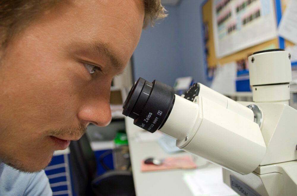
Biological samples play a crucial role in the field of microscopy, offering valuable insights into the intricate structures of living organisms. Before these samples can be examined under a microscope, they must undergo meticulous preparation to ensure clear and accurate imaging. The process of preparing biological samples for microscopy involves several steps that help maintain the integrity of the specimen and enhance the quality of the images obtained.
Sample Collection and Fixation
The first step in preparing biological samples for microscopy is the collection of the specimen. Whether it is a tissue sample, cell culture, or microorganism, proper collection techniques are essential to preserving the sample’s structure and characteristics. Once collected, the sample must be fixed to prevent degradation and maintain its morphology. Fixation involves treating the sample with a chemical agent that stabilizes its structure by cross-linking proteins and nucleic acids. Common fixatives include formaldehyde, glutaraldehyde, and paraformaldehyde, each with specific applications based on the type of sample being prepared.
Dehydration and Clearing
After fixation, biological samples are typically dehydrated to remove water content and prepare them for embedding in a solid medium. Dehydration is achieved through a series of alcohol washes of increasing concentration, gradually replacing water within the sample with alcohol. Following dehydration, samples may undergo a clearing step to render them transparent and facilitate imaging. Clearing agents such as xylene or benzyl alcohol are used to remove residual alcohol and enhance the transparency of the sample for microscopy.
Embedding and Sectioning
Once dehydrated and cleared, biological samples are embedded in a solid medium to provide structural support during sectioning. Embedding involves infiltrating the sample with a material that solidifies to form a block, ensuring the sample remains intact and can be sliced into thin sections for microscopy. Common embedding media include paraffin wax for light microscopy or resin for electron microscopy, chosen based on the specific requirements of the imaging technique. Following embedding, samples are sectioned using a microtome, a precision instrument that cuts thin slices of the sample for mounting on microscope slides.
Staining and Mounting
Staining is a crucial step in preparing biological samples for microscopy, enhancing the contrast and visibility of cellular structures. Various dyes and stains are used to highlight specific components within the sample, such as nuclei, cytoplasm, or organelles. Staining techniques range from simple stains that color the entire sample to complex methods targeting specific cellular components. Once stained, samples are mounted on microscope slides using mounting media to secure the specimen and protect it from damage during imaging.
Imaging and Analysis
With the biological sample prepared and mounted on a slide, it is ready for microscopy. Microscopes equipped with different imaging techniques, such as brightfield, phase contrast, or fluorescence microscopy, allow researchers to visualize the intricate details of the sample at various magnifications. Advanced imaging technologies, such as confocal microscopy or electron microscopy, provide even higher resolution images for detailed analysis of cellular structures. Following imaging, the obtained data can be analyzed using digital image processing software to enhance contrast, remove noise, and generate detailed images for further study.
Future Perspectives in Sample Preparation
As microscopy techniques continue to advance, the field of biological sample preparation is also evolving to meet the demands of high-resolution imaging and analysis. Emerging technologies, such as 3D imaging and super-resolution microscopy, are pushing the boundaries of sample preparation methods to achieve unprecedented levels of detail in biological samples. Additionally, the integration of automation and robotics in sample preparation workflows is streamlining the process, reducing human error, and increasing throughput for large-scale studies. By staying abreast of these developments and continually refining sample preparation techniques, researchers can unlock new insights into the complex world of biological structures and functions.
In conclusion, the preparation of biological samples for microscopy is a meticulous process that involves multiple steps to ensure the integrity and quality of the specimen. From sample collection and fixation to staining, imaging, and analysis, each stage plays a critical role in obtaining clear and detailed images for scientific study. By following established protocols and embracing innovative technologies, researchers can uncover the hidden mysteries of the microscopic world and advance our understanding of living organisms at the cellular level.
